anatomical model
Showing 13–24 of 54 results
-
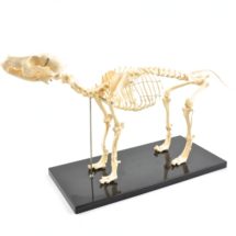
dog skeleton – removable model
MA699
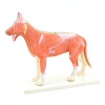
– one half of the body is presented with the coat, the other with the muscles and the tendons Ability to remove lung, heart, liver, stomach and intestine – possible opening of the rectum – hand painted – detailed representation of bones and spinal cord – dimensions: 36 x 15 x 34 cm – delivered on base -
dog skeleton – small model
-
dog skin disease
-
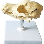
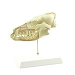
dog skull
-
dog skull anatomical model
-
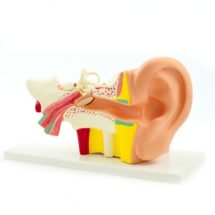
ears model – 4 parts
-
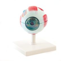
eye model – 6 parts
-
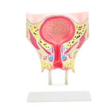
female bladder model
MA481
– about 15 cm high and mounted on a base. The anatomical model shows the most important structures of a female bladder and some of the surrounding life-size structures including: – muscle layer made of smooth muscle (Tunica muscularis) -squaters (Tunica mucosa) of the urothelium – structure and folds of the mucosa (Mucous folds) – bladder trine (Trigonum vesicae) – anastomosis of the ureter (Ostium ureteris) with inter-ureteral folds – urinary tract – urethra with Skene glands – vesical venous plexus (Plexus venosus vesicalis) – bulbus vestibuli (Vestibular bulb) – vaginal opening (Ostium vaginae) – in addition: lipid, bone and muscles -
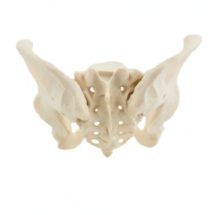
female pelvis – real size model
MA494
– sacrum (Os sacrum) – sacrum-ilion joint (sacroiliac joint) – Coxal bone (iliac crest) – ilium (Iliac bone) – anterior iliac bone (anterior iliac spine) – acetabulum – ischium (bone ischium) – ischiopharic foramina – pubis (pubic bone) with pubic arch (pubic arch) and symphysis pubis (symphysis) -
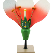
flower anatomy
-
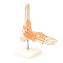
foot model
-
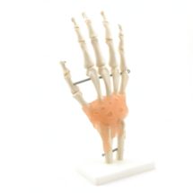
hand skeleton with ligaments


